Optics In Medicine And Life Sciences
The development and application of optics has helped modern medicine and life sciences enter a stage of rapid development, such as minimally invasive surgery, laser therapy, disease diagnosis, biological research, DNA analysis, etc.
Surgery and Pharmacokinetics
The role of optics in surgery and pharmacokinetics is mainly manifested in two aspects: laser and in vivo illumination and imaging.
1. Application of laser as energy source
The concept of laser therapy was introduced into eye surgery in the 1960s. When the different types of lasers and their properties were recognized, laser therapy was rapidly expanded to other fields.
Different laser light sources (gas, solid, etc.) can emit pulsed lasers (Pulsed Lasers) and continuous lasers (Continuous wave), which have different effects on different tissues of the human body. These light sources mainly include: pulsed ruby laser (Pulsed ruby laser); continuous argon ion laser (CW argon ion laser); continuous carbon dioxide laser (CW CO2); yttrium aluminum garnet (Nd:YAG) laser. Because continuous carbon dioxide laser and yttrium aluminum garnet laser have blood coagulation effect when cutting human tissue, they are most widely used in general surgery.
The wavelength of lasers used in medical treatment is generally greater than 100 nm. The absorption of lasers of different wavelengths in different tissues of the human body is used to expand its medical applications. For example, when the wavelength of the laser is greater than 1um, water is the primary absorber. Lasers can not only produce thermal effects in human tissue absorption for surgical cutting and coagulation, but also produce mechanical effects.
Especially after people discovered the nonlinear mechanical effects of lasers, such as the generation of cavitation bubbles and pressure waves, lasers were applied to photodisruption techniques, such as cataract surgery and kidney stone crushing chemical surgery. Lasers can also produce photochemical effects to guide cancer drugs with photosensitive mediators to release drug effects on specific tissue areas, such as PDT therapy. Laser combined with pharmacokinetics plays a very important role in the field of precision medicine.
2. The use of light as a tool for in vivo illumination and imaging
Since the 1990s, CCD (Charge-Coupled Device) camera was introduced into minimally invasive surgery (Minimally Invasive Therapy, MIT), and optics had a qualitative change in surgical applications. The imaging effects of light in minimally invasive and open surgery mainly include endoscopes, micro-imaging systems, and surgical holographic imaging.
Flexible Endoscope, including gastroenteroscope, duodenoscope, colonoscope, angioscope, etc.
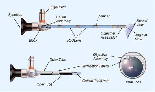
The optical path of the endoscope
The optical path of the endoscope includes two independent and coordinated systems of illumination and imaging.
Rigid Endoscope, including arthroscopy, laparoscopy, thoracoscopy, ventriculoscopy, hysteroscopy, cystoscopy, otolinoscopy, etc.
Rigid endoscopes generally only have several fixed optical path angles to choose from, such as 30 degrees, 45 degrees, 60 degrees, etc.
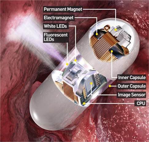
A miniature body camera is an imaging device based on a miniature CMOS and CCD technology platform. For example, a capsule endoscope,PillCam. It can enter the digestive system of the human body to check for lesions and monitor the effects of drugs.
The capsule endoscope
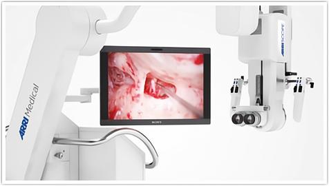
Surgical holographic microscope, an imaging device used to observe 3D images of fine tissue in precision surgery, such as neurosurgery for craniotomy.
The surgical holographic microscope
Summarize:
1. Due to the thermal effect, mechanical effect, photosensitivity effect and other biological effects of the laser, it is widely used as an energy source in minimally invasive surgery, non-invasive treatment and targeted drug therapy.
2. Due to the development of imaging technology, medical optical imaging equipment has made great progress in the direction of high resolution and miniaturization, laying the foundation for minimally invasive and precise surgery in vivo. At present, the most commonly used medical imaging devices include endoscopes, holographic images and micro-imaging systems.


